image
في هذه الصفحة سوف تجد مواضيع عن human anatomy lower abdomen وpicture of human body showing all organs، بالإضافة إلى stomach any pancreas anatomy وanatomy stomach liver، كذلك Large Body Organs، علاوة على صفحات في reproductive part of our body، أيضا human body organs liver و organs of the human body diagram، بإلإضافة إلى pics of the human anatomy for kids و stomach liver anatomy، كما ستجد مواضيع تتحدث عن stomach human body diagram و anterior view of human body، إلى جانب صفحات عن human body with stomach urinary and reproductive organs و human stomach and chest anatomy، علاوة على pictures of the body with skin liver lungs and intestine و location of large intestine in human body، كذلك مقالات تتحدث عن human body intestent و where are the kidneys liver stomach pancreas large intestine located in the human body، ايضا صفحات في موضوع human body ear nose eyes و picture of urinary bladder of human body
فيما يلي صفحات متعلقة بكلمة البحث: image
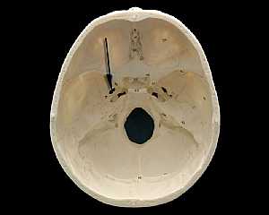
Skull Anatomy
19/10/2009 03:02:24 م
In This Section you will find detailed different Photos and images about the anatomy of the Skull bone including its surface , attachments related structures many more Items about the Skull anatomy... شاهد التفاصيل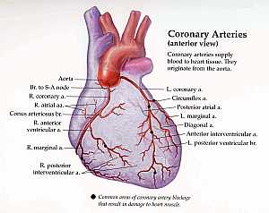
Coronary arteries anatomy
20/12/2009 02:25:00 م
this image shows the coronary arteries of the heart ( the arteries that supply the heart muscle with oxygen and nutrients) From anterior view .these arteries when occluded paretially or completely it... شاهد التفاصيل
Atlas of Human Anatomy
27/04/2006
In This Section you will find detailed different sections about all different parts of the human body including head and neck , chest , abdomen , upper limbs , pelvis and lower limbs and many more Sec... شاهد التفاصيل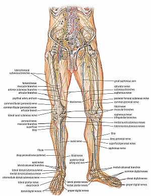
Leg nerves and vessels
15/07/2010 03:22:46 م
In This Section you will find detailed different Photos and images about the anatomy of the Leg nerves and vessels including parts , related structures and many more Items about the Leg nerves and ves... شاهد التفاصيل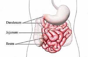
Spinal cord anatomy
22/10/2009 01:52:10 م
In This Section you will find detailed different Photos and images about the anatomy of the Spinal cord including its surface , parts , related structures , Functions of the spinal; cord , Spinal nerv... شاهد التفاصيل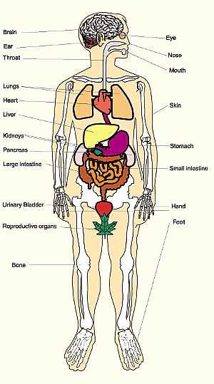
Anatomy of human body
12/11/2009 01:35:00 م
this image shows the human body from anterior view showing some organs of it showing: 1. brain 2. eye 3. nose 4. mouth 5. ear 6. throat 7. skin 8. lungs 9. heart 10. liver 11. kidneys 12. pancreas 13... شاهد التفاصيل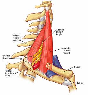
Neck Anatomy
19/10/2009 03:05:59 م
In This Section you will find detailed different Photos and images about the anatomy of the Neck including its surface , attachments , structures , Neck arteries , Neck Veins , trachea , Esophagus an... شاهد التفاصيل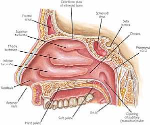
Nose anatomy
11/10/2009 03:55:00 م
this is a longitudinal section in the nasal cavity showing its medial wall showing: 1. frontal sinus 2. superior turbinate 3. middle turbinate 4. inferior turbinate 5. vestibule 6. hard palate 7. soft... شاهد التفاصيل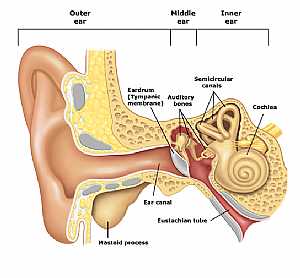
Ear anatomy
12/10/2009 04:42:00 ص
this shows the structure of the ear outer,middle and inner ear adjoined together showing: 1. mastoid process 2. ear canal 3. Eustachian tube 4. ear drum 5. auditory nerves 6. semicircular canals 7. co... شاهد التفاصيل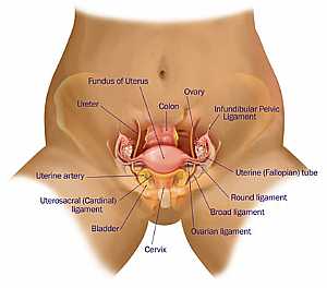
Female pelvic anatomy
13/11/2009 07:04:00 ص
this is an anterior view of the female reproductive system in the pelvic region showing: 1. fundus of the uterus 2. ureter 3. colon 4. ovary 5. infundibular pelvic ligament 6. uterine (fallopian ) tu... شاهد التفاصيل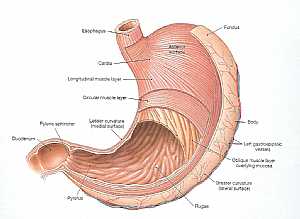
Stomach anatomy
09/12/2009 06:08:00 ص
this image shows the anatomy of the stomach showing its main features and parts.in this images we see the wall of the stomach being removed from the anterior portion to display the contents of the sto... شاهد التفاصيل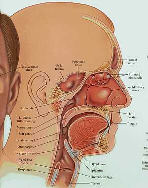
anatomy of the head and neck
09/10/2009 03:44:00 م
this image shows longitudinal section in the head and neck with their details (the frontal,nasal,oral and neck regions) showing: 1. nasolacrimal duct 2. sella turcica 3. sphenoid sinus 4. frontal sinu... شاهد التفاصيلCoronary arteries anatomy
20/12/2009 02:25:00 م
this image shows the coronary arteries of the heart ( the arteries that supply the heart muscle with oxygen and nutrients) From anterior view .these arteries when occluded paretially or completely it... شاهد التفاصيل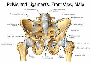
Bones and ligaments of the MALE Pelvis
22/11/2009 05:57:00 ص
this image shows the anterior view of the male pelvic brim ( the bones and ligaments that forms and supports the pelvic region) showing: 1. 4th lumbar vertebra 2. anterior sacroiliac ligament 3. ante... شاهد التفاصيل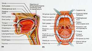
Oral Cavity
11/10/2009 04:09:00 م
the image on the right: is a detailed image of the structures of the mouth the image on the left: cut section in the head showing the structures of the mouth and oropharynx ( just after the mouth area... شاهد التفاصيل
Lung anatomy
16/07/2010 05:10:57 ص
In This Section you will find detailed different Photos and images about the anatomy of the Lung including its surface , parts , related structures , Lobes of the lungs , Hilum anatomy , alveolar anat... شاهد التفاصيل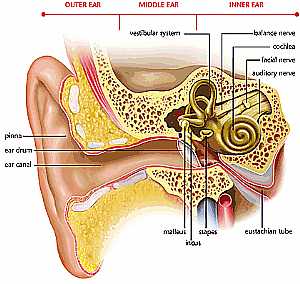
Ear anatomy
12/10/2009 04:21:00 ص
this image shows the different structures of the ear (external , middle and inner ear ) showing: 1. ear pinna 2. ear drum 3. ear canal 4. malleus 5. incus 6. stapes 7. eustacian tube 8. vestibular sy... شاهد التفاصيل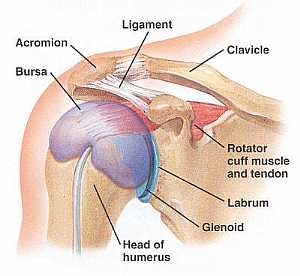
Shoulder joint anatomy
18/12/2009 07:48:00 ص
this image shows the anatomy of the shoulder joint from anterior view displaying the bones , ligaments and bursa of that joint. showing: 1. Acromion process of the scapula 2. Bursa of the shoulder jo... شاهد التفاصيل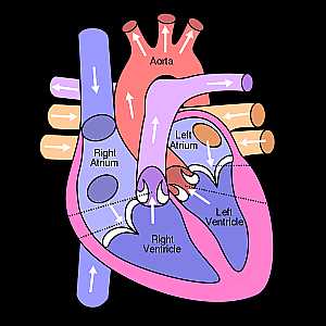
Heart anatomy
16/07/2010 05:10:26 ص
In This Section you will find detailed different Photos and images about the anatomy of the Heart including its surface , parts , related structures , Blood circulation of the heart , coronaries anato... شاهد التفاصيلFemale pelvic anatomy
13/11/2009 07:04:00 ص
this is an anterior view of the female reproductive system in the pelvic region showing: 1. fundus of the uterus 2. ureter 3. colon 4. ovary 5. infundibular pelvic ligament 6. uterine (fallopian ) tu... شاهد التفاصيلOral Cavity
11/10/2009 04:09:00 م
the image on the right: is a detailed image of the structures of the mouth the image on the left: cut section in the head showing the structures of the mouth and oropharynx ( just after the mouth area... شاهد التفاصيل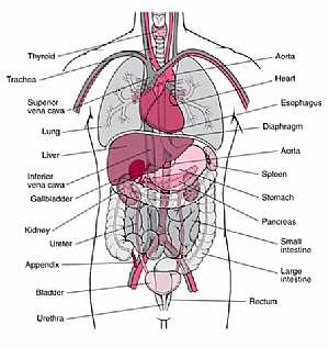
Human organs
05/03/2009 03:19:00 م
this image displays the important organs of the human body in the thorax , the abdomen and the pelvis showing: 1. thyroid gland 2. trachea 3. aorta 4. heart 5. esophagus 6. diaphragm 7. spleen 8. sto... شاهد التفاصيل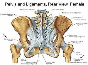
Bones and ligaments of the FEMALE Pelvis
22/11/2009 06:02:00 ص
this image shows the posterior "back" view of the female pelvic brim (the bones and ligaments that forms the pelvic region in the female) showing: 1. supraspinous ligament 2. posterior sacroi... شاهد التفاصيل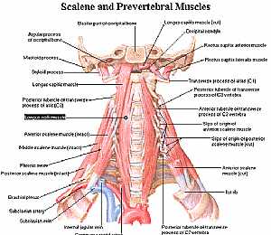
Neck Anatomy
11/10/2009 04:06:00 م
this is a back view of the muscles of the neck showing: 1. longus capitus m. 2. longus coli 3. ant. scalene m. 4. middle scalene m. 5. phrenic nerve 6. post. scalene m. 7. brachial plexus 8. subclavia... شاهد التفاصيل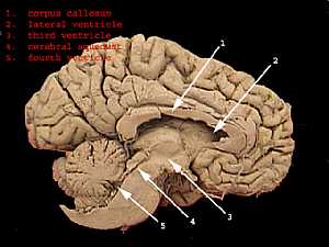
Brain ventricles
13/10/2009 01:20:00 ص
this section that shows the medial wall of the cerebral hemisphere shows the main four ventricles of the brain showing: 1. corpus callosum 2. lateral ventricle 3. cerebral aquiduct 4. fourth ventricle... شاهد التفاصيل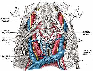
Neck anatomy
11/10/2009 04:02:00 م
this image shows the major vessels in the neck region and their relations to each other showing: 1. sternothyroid m. 2. inf. thyroid vein 3. middle thyroid vein 4. cricothyroid m. 5. sternothyroid art... شاهد التفاصيل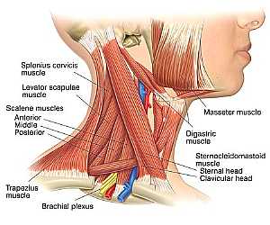
Neck Anatomy
11/10/2009 03:40:00 م
this is the muscles of the neck showing: 1. splenius cervicis m. 2. levator scapulae m. 3. masseter m. 4. digastric m. 5. sternocleidomastoid m. 6. brachial plexus 7. trapezius m. 8. ant. , middle and... شاهد التفاصيلShoulder Anatomy
14/07/2010 11:39:39 ص
In This Section you will find detailed different Photos and images about the anatomy of the Shoulder including its surface , parts , related structures , Muscles of the shoulder , Brachial plexus , te... شاهد التفاصيل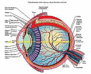
Eye anatomy
11/10/2009 04:00:00 م
this diagram details the different parts and structures of the human eye showing: 1. conjunctiva 2. ora serrata 3. cilliary body 4. aqueous 5. iris 6. ant. chamber 7. cornea 8. pupil 9. lens 10. post.... شاهد التفاصيل
Cranial nerves anatomy
05/11/2009 04:03:00 ص
this image shows the all cranial nerves and displaying their effector organs showing: 1. Olfactory nerve I 2. Optic nerve II 3. Occulomotor nerve III 4. Trochlear nerve IV 5. Trigeminal nerve V 6. Ab... شاهد التفاصيل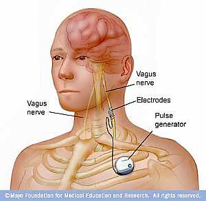
Vagus nerve anatomy
01/11/2009 02:28:00 م
this image shows the course of the vagus nerve descending from the brain to the heart ( attached to it the pulse generator "pace maker")... شاهد التفاصيل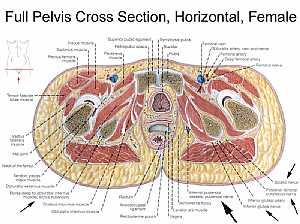
Female pelvic anatomy
13/11/2009 07:31:00 ص
this image is a horizontal section in a female pelvis shows the different pelvic organs and structures in relation to each other showing: 1. iliacus muscle 2. sartorius muscle 3. rectus femoris muscl... شاهد التفاصيلCranial nerves anatomy
22/10/2009 01:51:53 م
In This Section you will find detailed different Photos and images about the anatomy of the Cranial Nerves including Their types , Fascial nerve anatomy , trigeminal nerve anatomy , vagus nerve anatom... شاهد التفاصيل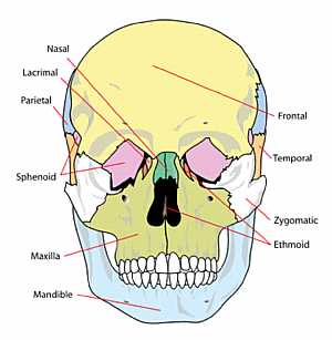
Skull Anatomy
10/10/2009 04:26:00 م
this image shows the names of the bones that forms the front of the skull showing: 1. nasal bone 2. lacrimal bone 3. temporal bone 4. zygomatic bone 5. ethmoid bone 6. mandible bone 7. sphenoid bone 8... شاهد التفاصيل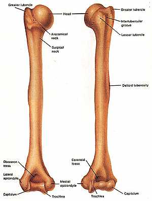
Humerus bone
04/12/2009 06:27:00 ص
this image shows the humerus bone (the bone of the arm " links the shoulder to the elbow joint") from anterior and posterior view showing: 1. greater tubercle 2. head of the humerus 3. anatom... شاهد التفاصيل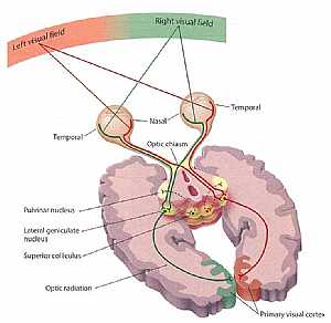
Visual pathway
03/11/2009 03:29:00 م
this image shows the visual pathway that carry sensation form the eye to the cerebral cortex showing: 1. temporal and nasal retina 2. optic nerve 3. optic chiasm 4. optic tract 5. pulvinar nucleus 6.... شاهد التفاصيل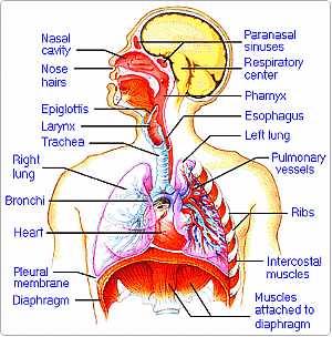
Respiratory system
27/04/2006
<b>The Respiratory System - Glossary <-b> <b>Bronchi<-b>: The two main air passages into the lungs. <b>Diaphragm<-b>: The main muscle used for breathing; separates the chest cavity from the abdomin... شاهد التفاصيلSpinal cord anatomy
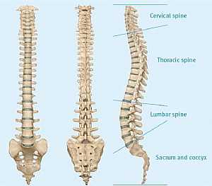
23/10/2009 03:51:00 م
this shows the different segments of the vertebral column showing: 1. cervical 2. thoracic 3. lumbar 4. sacral 5. coccygeal... شاهد التفاصيل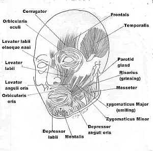
Anatomy of the face
10/10/2009 04:23:00 م
this is a detailed image for the fascial muscles ( the muscles of the face) showing: 1.frontalis m. 2. temporalis m. 3. parotid gland 4. risorius m. 5. masseter m. 6. zygomaticus major m. 7. zygomatic... شاهد التفاصيل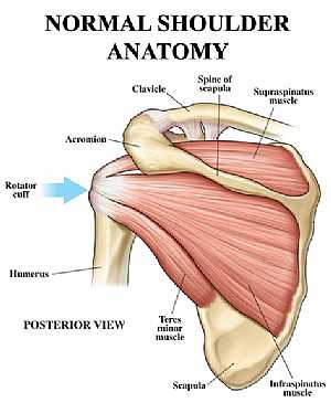
Shoulder joint anatomy
18/12/2009 07:20:00 ص
this image shows the anatomy of the shoulder joint from posterior view displaying the bones, tendons and muscles of the joint in relation to each other. showing: 1. Supraspinatus muscle 2. Spine of t... شاهد التفاصيل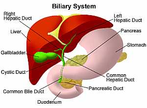
Anatomy of the liver"biliary system"
05/03/2009 04:40:00 م
this image shows the biliary system ( with the liver and the stomach) showing: 1. right hepatic duct 2. liver 3. gall bladder 4. cystic duct 5. common bile duct 6. duodenum 7. pancreatic duct 8. comm... شاهد التفاصيل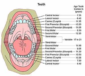
Anatomy of the mouth
11/10/2009 03:45:00 م
this image shows the names of the teeth in the mouth showing: 1. incisors 2. canines 3. premolars 4. molars... شاهد التفاصيل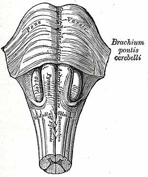
Brain stem anatomy
22/10/2009 01:50:57 م
In This Section you will find detailed different Photos and images about the anatomy of the Brain Stem including its surface , parts , related structures , midbrain anatomy , pons anatomy , medulla an... شاهد التفاصيل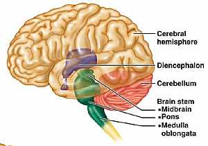
Brain anatomy
13/10/2009 12:39:00 ص
this is lateral view of the brain showing the cerebrum, cerebellum and their relation to the diencephalon (hypothalamus) and the brain stem (the root of the brain) showing: 1. cerebral hemisphere 2. d... شاهد التفاصيل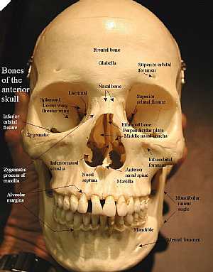
Anterior skull anatomy
10/10/2009 04:32:00 م
detailed image for the front of the skull showing: 1. frontal bone 2. glabella 3. nasal bone 4. superior orbital foramen 5. superior orbital fissure 6. ethmoid bone 7. infraorbital foramen 8. ant. nas... شاهد التفاصيل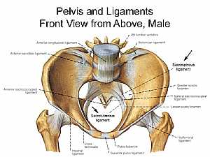
Bones and ligaments of the MALE Pelvis
22/11/2009 05:51:00 ص
this image shows an superior view of the MALE pelvic brim (bones and ligaments that forms and surrounds the pelvic region) showing: 1. anterior longitudinal ligament 2. anterior sacroiliac ligament 3... شاهد التفاصيل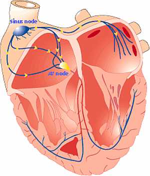
Natural pacemaker of the heart
20/12/2009 02:19:00 م
this image shows the natural passage of the nerve impulses in the cardiac muscle.This image shows the natural pacemaker of the heart (SA node)"sinoatrial node" that sinus has its own rhythmic... شاهد التفاصيل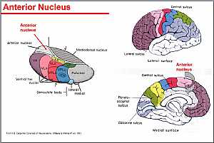
Thalamus anatomy
22/10/2009 02:21:00 م
this image shows the details of the thalamus and its nuclei showing: 1. anterior nuclei 2. ventral nuclei 3. geniculate body 4. lateral nuclei 5. medial nuclei 6. mediodorsal nucleus... شاهد التفاصيل
Neck Anatomy
11/10/2009 03:40:00 م
this is the muscles of the neck showing: 1. splenius cervicis m. 2. levator scapulae m. 3. masseter m. 4. digastric m. 5. sternocleidomastoid m. 6. brachial plexus 7. trapezius m. 8. ant. , middle and... شاهد التفاصيل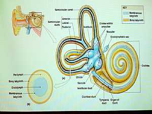
inner ear anatomy
12/10/2009 04:19:00 ص
this is a detailed image for the inner ear showing: 1. semicircular canals 2. semicircular ducts ( ant , post and middle) 3. vestibule 4. ampullae 5. maculae 6. endolymphatic sac 7. utricle 8. saccule... شاهد التفاصيل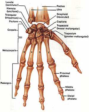
Hand anatomy
04/12/2009 06:23:00 ص
this image shows the anatomy of the hand displaying the bones forming the hand (carpal , metacarpal , phalanges bones) showing: 1. radius bone 2. ulna bone 3. scaphoid bone 4. capitate bone 5. trapez... شاهد التفاصيل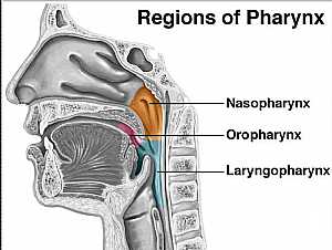
Pharynx anatomy
12/10/2009 04:34:00 ص
this image explains the position of the three parts of the pharynx showing: 1. nasopharynx 2. oropharynx 3. laryngopharynx... شاهد التفاصيل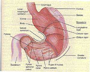
Stomach anatomy
09/12/2009 06:09:00 ص
this image shows the anatomy of the stomach showing its main features and parts.in this images we see the wall of the stomach being removed from the anterior portion to display the contents of the sto... شاهد التفاصيل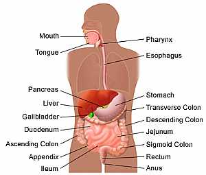
Systemic anatomy of the human body
11/11/2009 03:03:45 م
In This Section you will find detailed different Photos and images about the Different Systems of the human body including Nervous system , The circulatory system , Skeletal System and many more Item... شاهد التفاصيل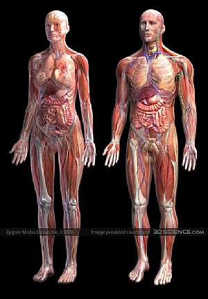
Muscles Anatomy (muscular system)
05/03/2009 02:10:00 م
this image shows the different muscles the forms and supports our human body and differs between male and female... شاهد التفاصيل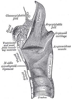
Larynx Anatomy
19/10/2009 03:06:37 م
In This Section you will find detailed different Photos and images about the anatomy of the Larynx including its surface , attachments , related structures , vocal cords and many more Items about the... شاهد التفاصيلالاستمناء قبل التحلل في العمرة
, , , , , , , ,هل يجوزتأخير فدية المحضور في العمرة
, , , , , , , ,ماهي وظائف المكبس بمحرك السيارة
, , , , , , , , , , , , , , , , , , , , ,هل يصح ارتداء شنطة مخيطة بالعمرة
, , , , , , , , , , , , ,