bone anatomy
في هذه الصفحة سوف تجد مواضيع عن human pelvis anatomy bones وanatomy body bones، بالإضافة إلى anatomy of pelvis وhuman hand anatomy bones، كذلك anatomy of bones chest، علاوة على صفحات في bone anatomy of the forearm، أيضا pelvic bone lateral view و free human anatomy bone chart، بإلإضافة إلى human bone anatomy chest و pelvic muscle anatomy، كما ستجد مواضيع تتحدث عن human anatomy diagram pelvic و sacrum bones muscles، إلى جانب صفحات عن pelvic joint anatomy و bone of pelvis، علاوة على pelvic phot و PEVIC BONES، كذلك مقالات تتحدث عن human pelvic anatomy diagram و anatomy of the pelvic bones، ايضا صفحات في موضوع pictures of human pelvic bones و diagram of pelvis bone
فيما يلي صفحات متعلقة بكلمة البحث: bone anatomy
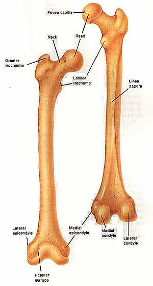
Femur bone anatomy
04/12/2009 07:30:00 ص
this image shows the anatomy of the femur bone from anterior and posterior view displaying its different features and parts showing: 1. fovea capitis 2. head of the femur 3. neck of the femur 4. less... شاهد التفاصيل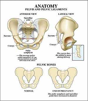
Pelvic bone anatomy
13/11/2009 06:32:00 ص
this image shows the pelvic bone that supports the pelvic region from anterior and lateral view showing: 1. sacroiliac joint 2. sacrum 3. coccyx 4. symphysis pubis 5. hib bone... شاهد التفاصيلFemur bone anatomy
04/12/2009 07:30:00 ص
this image shows the anatomy of the femur bone from anterior and posterior view displaying its different features and parts showing: 1. fovea capitis 2. head of the femur 3. neck of the femur 4. less... شاهد التفاصيل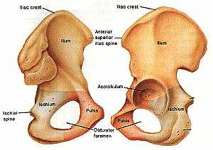
Hib bone anatomy
04/12/2009 07:22:00 ص
this image shows the anatomy of the hib bone from anterio-lateral (on the left) and from posterio-lateral view(on the right) showing: 1. iliac crest 2. ilium bone 3. ischial bone 4. pubis 5. obturato... شاهد التفاصيلHib bone anatomy
04/12/2009 07:22:00 ص
this image shows the anatomy of the hib bone from anterio-lateral (on the left) and from posterio-lateral view(on the right) showing: 1. iliac crest 2. ilium bone 3. ischial bone 4. pubis 5. obturato... شاهد التفاصيل
Skull Anatomy
19/10/2009 03:02:24 م
In This Section you will find detailed different Photos and images about the anatomy of the Skull bone including its surface , attachments related structures many more Items about the Skull anatomy... شاهد التفاصيل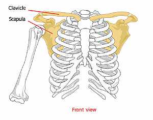
Pectoral anatomy
04/12/2009 06:50:00 ص
this image shows the bones forming the pectoral girdle from anterior view (the clavicle and scapula bone) displaying their position and relation to the rest of the body bones... شاهد التفاصيل
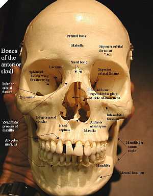
Anterior skull anatomy
10/10/2009 04:32:00 م
detailed image for the front of the skull showing: 1. frontal bone 2. glabella 3. nasal bone 4. superior orbital foramen 5. superior orbital fissure 6. ethmoid bone 7. infraorbital foramen 8. ant. nas... شاهد التفاصيل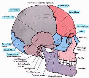
Skull Anatomy
10/10/2009 04:28:00 م
this shows the names of the bones that forms the side of the skull showing: 1. coronal suture 2. frontal bone 3. sphenoid bone 4. orbital bone 5. nasal bone 6. zygomatic bone 7. maxillary bone 8. mand... شاهد التفاصيل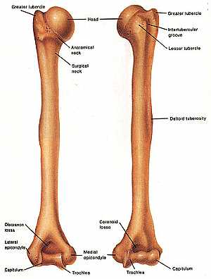
Humerus bone
04/12/2009 06:27:00 ص
this image shows the humerus bone (the bone of the arm " links the shoulder to the elbow joint") from anterior and posterior view showing: 1. greater tubercle 2. head of the humerus 3. anatom... شاهد التفاصيل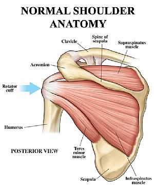
Shoulder joint anatomy
18/12/2009 07:20:00 ص
this image shows the anatomy of the shoulder joint from posterior view displaying the bones, tendons and muscles of the joint in relation to each other. showing: 1. Supraspinatus muscle 2. Spine of t... شاهد التفاصيل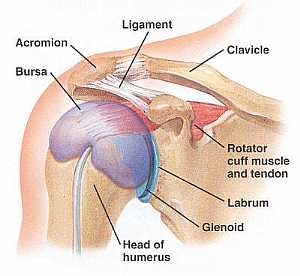
Shoulder Anatomy
14/07/2010 11:39:39 ص
In This Section you will find detailed different Photos and images about the anatomy of the Shoulder including its surface , parts , related structures , Muscles of the shoulder , Brachial plexus , te... شاهد التفاصيل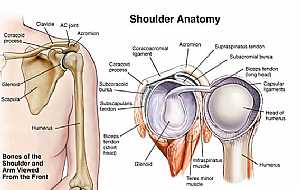
Shoulder joint anatomy
18/12/2009 07:45:00 ص
This image shows the anatomy of the shoulder joint from anterior view (on the left) and an open view (on the right) showing: 1. Supraspinatus tendon 2. Subacromial bursa 3. Biceps tendon (long head) ... شاهد التفاصيل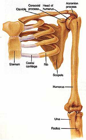
Shoulder joint anatomy
04/12/2009 07:07:00 ص
this image shows the shoulder joint displaying the bones and ligaments that form the joint and supports it (from anterior view) showing: 1. clavicle bone 2. scapula bone 3. humerus bone... شاهد التفاصيل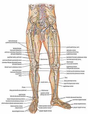
Nerves of the lower limb anatomy
24/11/2009 06:08:00 ص
this image shows the nerves of the lower limb showing their course , relation , branches and distribution (from anterior view) showing: "numbers" abdomen: 1. hepatic plexus 2. L1 nerve 3. L2... شاهد التفاصيل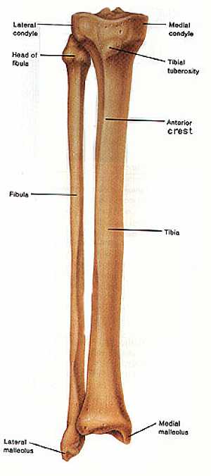
Fibula and Tibia bones anatomy
04/12/2009 07:43:00 ص
this image shows the anatomy of the fibula and tibia bones (the bones of the leg) in relation to each other ,displaying the different features and parts of them ,fibula (on the left) and the tibia (on... شاهد التفاصيل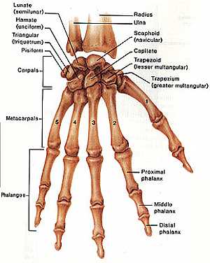
Hand anatomy
04/12/2009 06:23:00 ص
this image shows the anatomy of the hand displaying the bones forming the hand (carpal , metacarpal , phalanges bones) showing: 1. radius bone 2. ulna bone 3. scaphoid bone 4. capitate bone 5. trapez... شاهد التفاصيل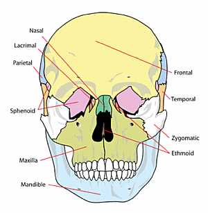
Skull Anatomy
10/10/2009 04:26:00 م
this image shows the names of the bones that forms the front of the skull showing: 1. nasal bone 2. lacrimal bone 3. temporal bone 4. zygomatic bone 5. ethmoid bone 6. mandible bone 7. sphenoid bone 8... شاهد التفاصيل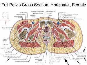
Female pelvic anatomy
13/11/2009 07:31:00 ص
this image is a horizontal section in a female pelvis shows the different pelvic organs and structures in relation to each other showing: 1. iliacus muscle 2. sartorius muscle 3. rectus femoris muscl... شاهد التفاصيل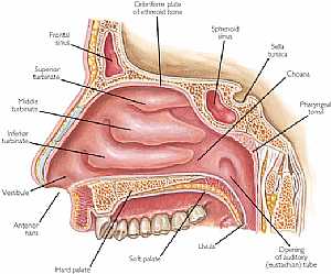
Nose anatomy
11/10/2009 03:55:00 م
this is a longitudinal section in the nasal cavity showing its medial wall showing: 1. frontal sinus 2. superior turbinate 3. middle turbinate 4. inferior turbinate 5. vestibule 6. hard palate 7. soft... شاهد التفاصيلAnterior skull anatomy
10/10/2009 04:32:00 م
detailed image for the front of the skull showing: 1. frontal bone 2. glabella 3. nasal bone 4. superior orbital foramen 5. superior orbital fissure 6. ethmoid bone 7. infraorbital foramen 8. ant. nas... شاهد التفاصيلSkull Anatomy
10/10/2009 04:26:00 م
this image shows the names of the bones that forms the front of the skull showing: 1. nasal bone 2. lacrimal bone 3. temporal bone 4. zygomatic bone 5. ethmoid bone 6. mandible bone 7. sphenoid bone 8... شاهد التفاصيلالاستمناء قبل التحلل في العمرة
, , , , , , , ,هل يجوزتأخير فدية المحضور في العمرة
, , , , , , , ,ماهي وظائف المكبس بمحرك السيارة
, , , , , , , , , , , , , , , , , , , , ,هل يصح ارتداء شنطة مخيطة بالعمرة
, , , , , , , , , , , , ,