hypoglossal nerve
في هذه الصفحة سوف تجد مواضيع عن medical pictures of the skull وmajor nerves of the skull، بالإضافة إلى hypoglossal canal of the skull وbase view of the skull، كذلك BASE OF HUMAN SKULL، علاوة على صفحات في bottom of skull، أيضا base of skull anatomy و base of skull، بإلإضافة إلى what muscles are at the base of skull و image of skull base from above، كما ستجد مواضيع تتحدث عن hypoglossal nerve و base of the skull، إلى جانب صفحات عن base of skull nerves و mylohyoid muscle، علاوة على hypoglossal nerve و human nerve anatomy diagram، كذلك مقالات تتحدث عن lingual nerve anatomy و mylohyoid muscle، ايضا صفحات في موضوع picture mylohyoid muscle و hypoglossal nerve
فيما يلي صفحات متعلقة بكلمة البحث: hypoglossal nerve
Hypoglossal nerve anatomy
03/11/2009 02:53:00 م
this image shows the hypoglossal nerve at its origin in relation to the surrounding structures showing: 1. hypoglossal nerve 2. cranial root of spinal accessory nerve 3. spinal root of spinal accesso... شاهد التفاصيل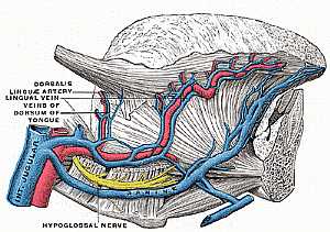
Hypoglossal nerve anatomy
03/11/2009 02:15:00 م
this image shows the hypoglossal nerve in the region just under the tongue in relation to the surrounding structures showing: 1. hypoglossal nerve 2. dorsalis muscle 3. lingual nerve 4. veins of the... شاهد التفاصيل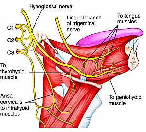
Hypoglossal nerve anatomy
03/11/2009 02:13:00 م
this image shows the cranial nerve XII "hypoglossal nerve" in the face region in the lateral aspect in relation to the surrounding structures showing: 1. hypoglossal nerve 2. lingual branch o... شاهد التفاصيل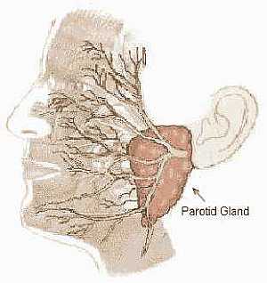
Fascial Nerve anatomy
30/10/2009 03:32:00 م
this image shows the position of the point of branching of the fascial nerve at the parotid gland... شاهد التفاصيل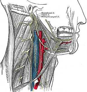
Cranial nerves X,VII and IX anatomy
25/10/2009 03:30:00 م
this image shows the course of the hypoglossal,vagus and glossopharygeal nerves showing: 1. hypoglossal nerve 2. vagus nerve 3. glossopharygeal nerve 4. lingual nerve 5. cervical branch... شاهد التفاصيل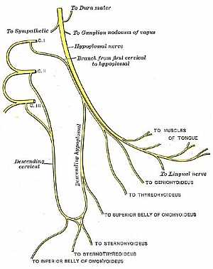
Hypoglossal nerve anatomy
03/11/2009 02:20:00 م
this diagram illustrates the hypoglossal nerve's origin and branches showing: 1. Hypoglossal nerve 2. nerve to dura matter 3. nerve to ganglion nodosum of vagus 4. branch from first cervical to h... شاهد التفاصيلHypoglossal nerve anatomy
03/11/2009 02:13:00 م
this image shows the cranial nerve XII "hypoglossal nerve" in the face region in the lateral aspect in relation to the surrounding structures showing: 1. hypoglossal nerve 2. lingual branch o... شاهد التفاصيل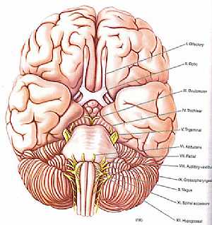
cranial nerves anatomy
14/10/2009 04:29:00 ص
a colored image for the cranial nerves exit showing: 1. olfactory nerve 2. optic nerve 3. occulomoto rnerve 4. trochlear nerve 5. trigeminal nerve 6. abducent nerve 7. fascial nerve 8. vestibulocochle... شاهد التفاصيل
Cranial nerves anatomy
22/10/2009 01:51:53 م
In This Section you will find detailed different Photos and images about the anatomy of the Cranial Nerves including Their types , Fascial nerve anatomy , trigeminal nerve anatomy , vagus nerve anatom... شاهد التفاصيل
Hypoglossal nerve anatomy
03/11/2009 02:53:00 م
this image shows the hypoglossal nerve at its origin in relation to the surrounding structures showing: 1. hypoglossal nerve 2. cranial root of spinal accessory nerve 3. spinal root of spinal accesso... شاهد التفاصيلHypoglossal nerve anatomy
03/11/2009 02:20:00 م
this diagram illustrates the hypoglossal nerve's origin and branches showing: 1. Hypoglossal nerve 2. nerve to dura matter 3. nerve to ganglion nodosum of vagus 4. branch from first cervical to h... شاهد التفاصيلHypoglossal nerve anatomy
03/11/2009 02:15:00 م
this image shows the hypoglossal nerve in the region just under the tongue in relation to the surrounding structures showing: 1. hypoglossal nerve 2. dorsalis muscle 3. lingual nerve 4. veins of the... شاهد التفاصيل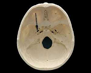
Skull Anatomy
19/10/2009 03:02:24 م
In This Section you will find detailed different Photos and images about the anatomy of the Skull bone including its surface , attachments related structures many more Items about the Skull anatomy... شاهد التفاصيل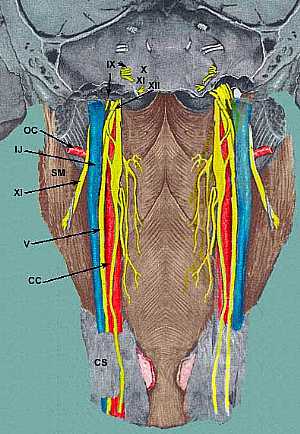
Cranial nerves anatomy
01/11/2009 02:27:00 م
this image shows the cranial nerves IX,X,XI,XII (glossopharyngeal , vagus , accessory and hypoglossal nerves) in relation to each other at the lateral aspect of the neck (pharynx) showing: 1. glossop... شاهد التفاصيل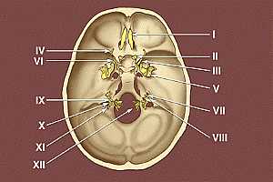
Base of the skull
09/10/2009 03:34:00 م
this image shows the exit of the twelve cranial nerves from the base of the skull * I. Olfactory nerve * II. Optic nerve * III. Oculomotor nerve * IV. Trochlear nerve * V. Trigeminal nerve * VI.... شاهد التفاصيل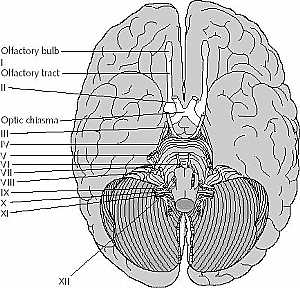
cranial nerves anatomy
14/10/2009 04:17:00 ص
this image is an inferior view of the brain showing the cranial nerves showing: 1. olfactory nerve 2. optic nerve 3. occulomoto rnerve 4. trochlear nerve 5. trigeminal nerve 6. abducent nerve 7. fasci... شاهد التفاصيل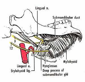
Hypoglossal nerve anatomy
30/10/2009 03:36:00 م
this image shows the hypoglossal nerve in relation to the surrounding structures just below the submandibular region showing: 1. lingual nerve 2. submandibular duct 3. mylohyoid muscle 4. mylohyoid m... شاهد التفاصيل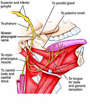
Glossopharybgeal nerve anatomy
30/10/2009 03:25:00 م
this image shows the glossopharyngeal nerve in the lateral aspect of the face displaying its course , branches and the related structures of the nerve showing: 1. superior ganglion 2. inferior gangli... شاهد التفاصيل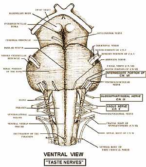
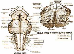
taste sensation pathway
02/11/2009 02:31:00 م
this image shows the origin of the nerves responsible for the taste sensation in relation to the surrounding structures of the brain stem showing: 1. intermediate portion of Cranial nerve VII "fa... شاهد التفاصيل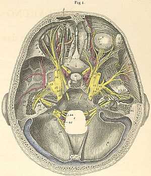
cranial nerves course in the skull
10/10/2009 03:09:00 م
this image shows the course of the cranial nerves inside the skull a) Frontal bone. b) Frontal sinus. c) Internal frontal spine. d) Foramen caecum. e) Crista galli. f) Frontal bone, orbital portion. g... شاهد التفاصيل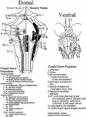
cranial nerves anatomy
14/10/2009 04:28:00 ص
this diagram shows location of the cranial nerves nuclei and their site of exit the right image is an anterior view and the left is a posterior view showing: 1. olfactory nerve 2. optic nerve 3. occul... شاهد التفاصيلGlossopharybgeal nerve anatomy
30/10/2009 03:25:00 م
this image shows the glossopharyngeal nerve in the lateral aspect of the face displaying its course , branches and the related structures of the nerve showing: 1. superior ganglion 2. inferior gangli... شاهد التفاصيل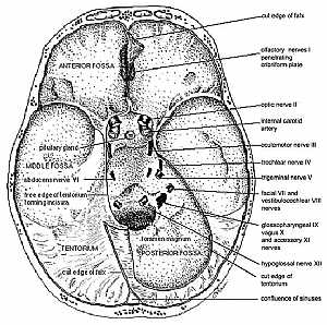
Intracranial Cavity
09/10/2009 12:13:00 م
section in the skull showing superior view of the base of the skull detailing the places where the cranial nerves exit (anterior,middle and posterior cranial fossae) showing: 1. olfactory nerve penetr... شاهد التفاصيل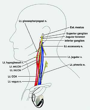
Glossopharybgeal nerve anatomy
29/10/2009 03:23:00 م
this is an image of the lateral side of the neck displaying the left glossopharyngeal nerve related to the surrounding structures showing: 1. left glossopharyngeal nerve 2. external auditory meatus 3... شاهد التفاصيلGlossopharybgeal nerve anatomy
29/10/2009 03:23:00 م
this is an image of the lateral side of the neck displaying the left glossopharyngeal nerve related to the surrounding structures showing: 1. left glossopharyngeal nerve 2. external auditory meatus 3... شاهد التفاصيلCranial nerves anatomy
05/11/2009 04:03:00 ص
this image shows the all cranial nerves and displaying their effector organs showing: 1. Olfactory nerve I 2. Optic nerve II 3. Occulomotor nerve III 4. Trochlear nerve IV 5. Trigeminal nerve V 6. Ab... شاهد التفاصيلcranial nerves anatomy
14/10/2009 04:17:00 ص
this image is an inferior view of the brain showing the cranial nerves showing: 1. olfactory nerve 2. optic nerve 3. occulomoto rnerve 4. trochlear nerve 5. trigeminal nerve 6. abducent nerve 7. fasci... شاهد التفاصيلالاستمناء قبل التحلل في العمرة
, , , , , , , ,هل يجوزتأخير فدية المحضور في العمرة
, , , , , , , ,ماهي وظائف المكبس بمحرك السيارة
, , , , , , , , , , , , , , , , , , , , ,هل يصح ارتداء شنطة مخيطة بالعمرة
, , , , , , , , , , , , ,